7 Point Plan for Mastitis Control

A Proposal

Over the past 40 years, since the adoption of the 5 Point Plan for Mastitis Control, there has been a significant reduction in the number of clinical cases of mastitis, in levels of bulk tank somatic cell counts and in the prevalence of mastitis caused by contagious bacteria.

Read more

Mastitis Basics

Mastitis Basics

Categories of Mastitis

Mastitis can be divided into:

  • Clinical and subclinical
  • Contagious and environmental
  • Lactation and dry period infections

Definitions

Clinical mastitis is when there are obvious changes to the udder tissue and / or the milk.  The milk may appear watery or full of clots, but will have an abnormal secretion.  The udder may appear normal or may be hot and swollen.  Depending on the severity of the infection, the cow may appear normal and behave accordingly or toxic mastitis can occur.

Subclinical mastitis is when there are no visible changes to the milk.  A milker will be unable to identify subclinical mastitis unless he carries out tests, such as individual cow cell counts, or the California mastitis test.  The cell count of quarters with subclinical mastitis will always be elevated above 200,000.

Contagious mastitis: the udder and teats are the reservoir of infection.  Transmission occurs during the milking process from udder preparation by contaminated hands, udder cloths and liners.  Infection establishes on the teat and teat canal.  Bacteria may then penetrate the mammary gland.  Most infections are subclinical and result in raised cell counts.  Control measures include post milking teat disinfection, dry cow therapy and culling.  Contagious bacteria include Staphylococcus aureus, Streptococcus agalactiae and dysgalactiae.  If your herd cell count is over 200,000 then this indicates that there is a problem with contagious mastitis.

Environmental mastitis: the environment is a reservoir of infection.  Infection is transmitted on to the teats between milkings or during udder preparation.  Organisms are forced up through the teat canal during the milking process or after milking if cows are allowed to lie down immediately following milking.  Most infections cause clinical mastitis.  Subclinical infections are less common.  Environmental mastitis is controlled by provision of a clean environment, adequate accommodation for cows, milking through a correctly functioning machine, good udder preparation and pre- milking teat disinfection.  Environmental organisms include E coli, Streptococcus uberis (straw bedding), Klebsiella (sawdust and shavings) and Bacillus.

Lactation period infection is when the bacteria enter the udder DURING lactation. For example this might be from spread of Staph aureus via the liner or an environmental infection of cows are lying in dirty beds.

Dry period infections are picked up when the cow is not lactating. These will almost always be environmental, E. coli and Strep uberis, as the risk of spread of contagious mastitis will be miniscule as the cow is not being milked.

QuestionsContagiousEnvironmental
BacteriaStaph aureus
Strep uberis
Strep agalactiae
Strep dysgalactiae
CNS
E.Coli
Strep uberis
Klebsiella
Pseudomonas
Source of infectionUdderEnvironment
Type of mastitis seenSub clinical all
Clinical [S. uberis & S. aureus]
Clinical & Sub clinical for Strep uberis only
Time of transmissionMilkingDry period
During milking
Between milking
Infection timeLactationLactation & dry period
Effect on herd cell countRaisedNone unless Strep uberis problems
Common control measuresDry cow therapy
Post milking teat dipping Culling
Treatment
Good hygiene during milking
Regular liner changes
Milking machine function
Clean environment
Internal teat seal
Clean udders and teats
Pre dipping
Milking machine function
Encouraging cows to stand after milking
Milking machine function
Vaccination

Benefits of thorough pre-milking routine

Benefits of thorough pre-milking routine


Although herdsmen may not have understood the biology of their pre-milking routine back in the days of milking in cow sheds, they knew that to not properly stimulate a cow lead to a more difficult hand milking and lower milk yields.  The invention of milking machines began to mask any downsides of not following a complete pre-milking routine.  With less labour on dairy farms and with increasing numbers of cows per person involved with the dairy herd, short cuts have been taken at milking time, with elements of an effective milking routine being removed – where they could not be automated.

Prime among these revolve around the pre-milking routine.  There was a trend – especially with the larger swing over milking parlours, to follow the philosophy of cows in, units on, cows out with very little if any teat preparation.  This was often based on the fact that cows’ teats were considered clean and milk hygiene results were not leading to a reduced milk price.  Where the somatic cell counts were high or clinical mastitis was an issue, the problem was very often blamed on the milking equipment or the housing.

However, in these situations dynamic milking assessments and observation of milk flow would indicate that the ineffective milking routine was leading to problems, and in many cases did not safe a great deal of time at milking.

Removing teat preparation from a milking routine, either throughout the year or when cows are turned out to graze ignores the substantial benefits that can be achieved.

Detecting abnormal milk or mastitis

Early detection of abnormal milk allows it to be excluded from the bulk tank, reducing bacterial counts and the somatic cell count (SCC). The early detection of abnormal milk also allows for early intervention and treatment which should reduce the recurrence rate and overall rate of new infections.

Stimulating milk let down

Manipulation of cows’ teats during pre-milking teat preparation results in milk let down.

A review of pre-milking teat preparation concluded that:

  • Less than 10 seconds of teat cleaning is not an adequate stimulus for consistent letdown response.
  • A teat cleaning procedure that results in a quality stimulus of 10 – 20 seconds is adequate to sanitise teats and achieve consistent milk letdown in most cows.
  • The optimum “window of time” to apply teat cups is 60 – 90 seconds after the cow’s teats and udder are first touched by the milker (this is known as the prep lag-time).
  • A prep-lag time of 60 seconds reduced average milking time per cow by 40 seconds and increased mean milk yield by 0.32kg per milking.

While the perception may be that milking can be speeded up by omitting pre-milking teat preparation, the opportunity to detect abnormal milk is lost and there are likely to be adverse effects on both the milking speed of cows and the amount of milk which they produce.  It is important that all milkers follow the same routine to optimise the benefits.

Milking cows is not just about throughput but the production of top quality milk that matches the demands of the market place.  Attention to detail nearly always pays.

Mastitis Detection

Mastitis Detection


Prompt and accurate mastitis detection is essential to ensure that clinical cases are treated successfully.

Clinical mastitis is inflammation of the udder where there are changes to the milk in the form of clots, abnormal colour or any other changes to the appearance of milk. These changes will be seen in all the milk. If clots are seen in the first two or three squirts and then the milk goes clear, this is NOT clinical mastitis but a local reaction in the teat itself. You do NOT need to carry out tests such as the California Mastitis Test (CMT) to decide if a cow has clinical mastitis. If the milk appears normal then the cow does NOT have clinical mastitis.

Early identification and treatment of mastitis will result in:

  • More rapid response to therapy
  • Reduce the risk of cross-infection, stopping other cows picking up mastitis
  • Stopping mastitis milk going into the bulk supply, protecting the bulk tank cell count and Bactoscan
  • Fewer repeat cases and so fewer cows being treated
  • Faster reduction in cell count
  • Faster return to milk production

Not every herd strips cows before milking. In some parts of the world the milking unit is applied without any teat preparation. Some herds may just strip the high risk group like the high yielding cows. In some herds mastitis detection is delayed as milkers may not foremilk or look for clinical signs and so any way that this can be improved will be an advantage.

In-line mastitis detectors have a number of advantages:

  • Pick up clots
  • Trap dirt or pieces of straw which helps focus the milker on improving teat preparation
  • Large detectors make it easy to see these clots
  • Large volume detectors, like the Ambic Vision, should not interfere with vacuum stability or milk flow provided they are kept clean


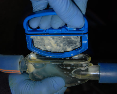
Clots captured on a Mastitis Detector Screen


Clots captured on a Mastitis Detector Screen

It is essential that milkers check the in-line detectors after every cow is milked. In recorder jar plants they should be checked before the milk is released to the bulk tank, as if this cow has mastitis her milk can then be discarded. If there are clots in a detector, then the cow should be examined for clinical mastitis. If in doubt, she can be rechecked at the next milking.

Be aware that more and more farmers are using an internal teat sealant along with dry cow therapy. It is not uncommon for cows to shed pieces of teat sealant for the first week after calving. These may well appear as clots in the detector, but they breakdown and have a distinct feel compared to mastitis clots.

It is NOT advisable to treat a cow on the basis that there are clots in the filter alone. The cow should be stripped to check for the presence of clinical mastitis. Remember that in-line detectors are an aid to mastitis detection, they will not necessarily pick up all cases.

Click here for further details on the Vision Mastitis Detector suitable for 16 – 19 mm milk tube.

Click here for further details on the Original Mastitis Detector suitable for 12 – 13 mm milk tube.